In the last week, three medical students have written to me asking the same thing: how to study pathological anatomy with memory techniques!
I understand them, because pathological anatomy is one of the most tragic examinations in medicine: long, complex, and full of data to memorize.
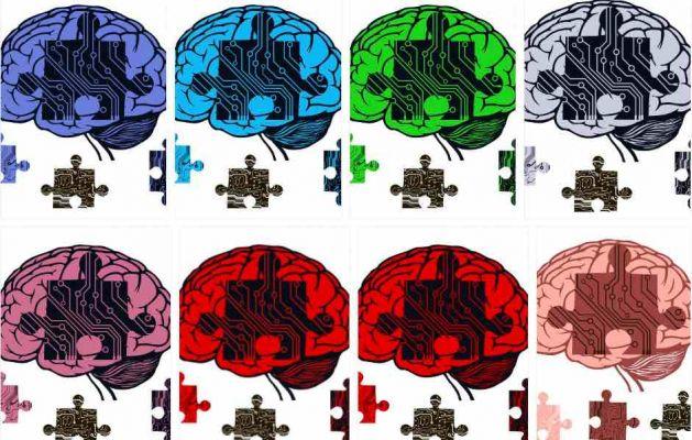
It is therefore an excellent example to talk about how to use 360-degree memory techniques, for any subject that is, in fact, difficult and long to memorize.
One of the three, Alex, is trying to use the memory palace, but is encountering some difficulties:
"Good morning,
I recently discovered your blog.
I am a medical student, and have found it difficult to use the Memory Building.
I tried to use it for the Pathological Anatomy exam, but the questions are many, each full of information
So I should be using a lot of Loci
Eg. Brain tumors:
1) There is an introductory part with various general information (epidemiology, age, complications, freezer exam)
2) Then the classification with 7 main types, and each type with its different subtypes.
Eg. Astrocytic tumors are divided into: astrocytomas, multiform glioblastomas, oligodendroglioma etc.
I need to know the characteristics of each subtype with any gradation (therefore another subdivision)
I have extreme difficulty because of this. I have read the various techniques (chain, Chinese box, locus) but I would need some ideas to organize my building well, perhaps with some examples.
Thank you very much, with you I discovered a world "
Answering Alex is therefore an excellent way not only to see how to study pathological anatomy with memory techniques, but also to address two fundamental points of the study with the techniques:
- How structured and effective memory palaces are built
- How to gradually start using memory techniques
In the first part of the article we will therefore see, with simple examples, the basics to really use memory techniques in the study.
In the second part you will find my answer to Alex, relating specifically to pathological anatomy, and structured on the basis of what was theoretically seen in the first.
So let's begin to see how to study pathological anatomy with memory techniques, clarifying the bases of their practical application.
3 ways to memorize a simple word list
Imagine you need to remember the following list of simple words:
Dog, belt, camel, sword, mouse, tree, patch, teapot, wheel, moon, umbrella, cat, canoe, bow, flag
You can repeat the 15 words to exhaustion, and maybe learn them in order in 5/10 minutes, only to have forgotten them an hour later.
Or, if you know memory techniques, you can learn them in a couple of minutes and still remember them well the next day.
And you can do it with three methods that have increasing complexity, but also increasing effectiveness.
Storage method 1: simple associations
It involves creating an image for each word and tying the images together with simple associative techniques.
If you don't know how, in the articles "remembering through images", "visual memory" and "phonetic conversion" you can see:
- Why converting to images is so memory efficient
- What should be the characteristics of the images you create
- How to start practicing with this list.
This first method of association is the very basis of memory techniques.
However, by itself, it is of little use for actual study.
Yet many try to study with memory techniques subjects such as private law, anatomy, analysis 1, macroeconomics ... without even mastering this first, simple, and basically not very useful, method.
It is simply impossible then that they can actually apply them in a complex study.
Memorization method 2: "linear" loci technique
A second method is to build a linear mnemonic path with the loci technique.
In this case, you first create a path of loci: that is, you identify an ordered series of physical places that you know well.
And then, to each of them, you attach one of the images that represent the words. .
In this way you create an association between a long-term memory, the locus, and a short-term memory, the word / image to remember.
And already in this form the method of the loci is quite useful, because:
- The association between long-term and short-term memory significantly improves recall
- You are able to remember the info even in no particular order, by going to the corresponding locus
And these two facts alone amply compensate for the effort and time spent building your Loci path.
But you are still a long way from using memory techniques effectively to study pathological anatomy, or any other complex subject. At the limit, you can take advantage of it here and there for some data list.
Method of memorization 3: technique of “segmented” loci, or memory palace.
A third method, which is a variant of the second, is that of the segmentation of the loci.
In this technique, on the main image you find details, usually 4 or 5, in a logical order.
These details will make you "menmonic hook”For subsequent images to remember, so that you will have a main image on which one or more secondary images are grouped.
So, for example, to memorize the 15 words from before, with the segmentation of the loci you have to proceed as follows:
- You create a path of only 3 loci. For example: lock on your front door, doormat, umbrella stand
- You visualize the first image corresponding to the first word, DOG, and associate it with the first locus, the lock. Imagine, for example, that your poor dog is stuck inside the lock, or that of a friend if you don't have one. If you see this absurd image vividly in your mind, trust me you will never forget it!
- Then, on the image of the dog, identify 4 points: for example, nose, eyes, ears, tail. It is important that the points follow a logical direction. In this case I went from the front of the dog to the back.
- At each point, which must be displayed very clearly, you then attach the next 4 words to memorize.
Then,
nose / belt
eyes / camel
ears / sword
tail / mouse
What you did, in a nutshell, was to use the info / image of the primary locus, DOG, as a reservoir for 4 secondary loci, to which you attached the secondary info, i.e. the next four words.
Proceeding in this way, on the second primary locus, doormat, you will attach the image of Tree, which is the sixth word.
And then, after segmenting the tree into 4 secondary loci, you will attach the next 4 words to memorize on each of them.
And so on.
In this way, you will have built a mini mnemonic tree, because your memory has two levels of structure:
A first level, which is that of the three keywords Dog-Tree-Umbrella.
And a second level that contains the others instead.
With the advantage, among other things, that you originally had to create a path of only 3 loci, instead of 15.
Let's say now that, after learning the 15 words with this third method, you have to learn 60 more ...
The simplest way is
- Take each of the 12 secondary images / concept you have already learned
- Divide it into 5 further loci, so as to have 60 in all.
- Associate the 60 new words with each of the new loci
You will thus have obtained a mnemonic tree with 3 distinct hierarchical levels.
And when these structures, with the multiplication of information, become very complex, you get what I call "palace of memory ".
Studying with concentric circle memory techniques
When you study with memory techniques you have to try to build structures of this type.
That is, you don't have to associate all the information one after the other with a single path of loci.
Also because it doesn't make sense from a conceptual point of view: in fact, the information is never all on the same level.
Knowledge of a subject, even when you study traditionally, presupposes instead of create hierarchies of information: primary, secondary, tertiary, and so on, depending on their importance and the logical connection between them.
And therefore, at each study cycle, you don't have to try to remember everything, but you have to proceed with the voluntary memorization in concentric circles.
Prima understanding and learning the main concepts, then the secondary ones, then the tertiaries, then the real details.
In this way:
- You have an exposure hierarchy that mirrors the conceptual importance hierarchy
- You first build a very solid overview, and then expand it by successive concentric circles
With or without memory techniques, this is the most effective way to study!
Now, maybe now you are a little perplexed, because the system seems quite complicated.
So take my word for it when I tell you it's extraordinarily effective and fast.
The problem, and it's certainly not your fault, is that you haven't practiced enough.
The problem of memory techniques
I have already spoken often about this aspect, among other things also recently, when answering a girl who wanted to use the techniques to study law.
The point is very simple: memory techniques, like any slightly sophisticated technique, take time to learn.
To explain what I mean, here are some examples:
- If you read a book on how to play tennis and then hit the court, you don't hit a ball and tennis seems very complicated to you.
- If you try to play pentatonic scales on the guitar after taking a guitar course of a few hours or even weeks, they will seem impossible.
- If they explain notes and fingering to you and then you start playing the piano, you won't be able to play almost anything
In short, it is normal that when a new activity requires the learning of a technique, at the beginning we carry it out in a slow, doubtful, awkward way.
And we don't get results until after considerable training.
While, I don't know why, many think they can use memory techniques effectively immediately after discovering them.
Memory techniques, on the other hand, are no exception to the exercise rule: to have results you must have the time, the will and the need to train.
And as with guitar or tennis, so it is with memory techniques: depending on how much effort and time you dedicate to them, you can be more or less good, and they will be more or less useful.
For this reason, I now report my answer to Alex, the medical student who has to prepare pathological anatomy.
Here you will find a practical example of the segmentation of the loci made before, and some advice to start using the techniques, starting in a simple way and then increasing the complexity.
How do you do for any technique!
How to study pathological anatomy
Hi Alex,
learning how to use memory techniques well takes some time, and starting with pathological anatomy is like having just started playing tennis and being up against Federer.
I try to explain myself as well as possible.
A mistake that many make is to start building a memory palace and try to attack too much information right away.
It is a too demanding "creative" effort of images and associations, which inevitably leads to great fatigue and discouragement.
All the more so if you have to study pathological anatomy, a descriptive subject full of details!
In fact, you have dozens and dozens of data for each page.
You must therefore proceed in a different way, which in my opinion is also the most correct way to study.
Concentric circles for pathological anatomy
Even when you study pathological anatomy in a traditional way, you certainly can't try to remember everything on the first or second reading.
Instead, you create a first set of very basic memories, and at each stage of study you expand them.
Also thanks to the fact that, having an overview, natural memory creates connections, reasoning, identifies similarities and differences. Allowing you to remember more, and with more sense!
You must proceed in the same way when you study with memory techniques: that is, a page should not be faced trying to remember it all, immediately, in a block.
It is impossible to do it with natural memory, and it is very hard to try to do it with memory techniques.
Instead, you have to do a first reading in which you build the backbone of the exam, distributing it in your memory building, with each pivotal element on a locus.
Then, with each rereading, you attach additional elements to that pivotal element.
That is, it is the same pivotal element that acts as a reservoir of loci for subsequent secondary information.
In this way you pass from a very precise overall view to subsequent detailed views.
A simple example for memorizing pathological anatomy
If you have to study brain tumors, first build a memory palace to remember the classification as it is: hard and pure. As if it were the index of a book.
Limiting yourself, at first reading, to actively memorize only ONE characteristic of the tumor in question, the one that you consider most important because it differentiates it from others.
Naturally, in addition to this voluntary and selective memorization, there will also be a first quota of involuntary and random memorization.
That is, you will remember a few things here and there, because they hit you, or because they are very logical, or for who knows what other reason.
At the next stage of study, you will then increase your voluntary memorization, building additional loci on those already built.
So let's say that you have memorized, on first reading, the entire classification of brain tumors, including the giant cell subependymal.
He is now in his good locus, to which the image of a SEGA is associated (It is the acronym with which it is identified!).
And you also actively memorized ONE main feature, which in this case by the way is already identified in the name (giant cell subependymal). Maybe thinking about a giant saw (double meanings are welcome, they help the memory!)
At this point, it is the image of the SEGA that becomes a reservoir of loci for the following info.
For example, divide it into handle, toothed part, non-toothed part.
The handle will therefore act as a locus for the association with the next info. And so on for the other parts.
Now, in this case it is easy because it is a rare tumor about which there is little to say, but it is right to explain the principle to you:
You have a first structure of loci to which you associate images / concept, and these concept images themselves become loci for the following info.
A more complex example
Let's now go to something more complex than the subependymal, for example diffuse astrocytoma.
Let's say that it is on your main path of loci, in particular in the locus identified by your bedside table, on which, to represent
diffuse astrocytoma, you have for example the image of the Milky Way (scattered stars = scattered stars….).
Here you have a first difficulty, because precisely, without a little practice, finding good images is not easy.
The Milky Way in my opinion is a discreet image, also because it can be further segmented, and therefore it is an excellent reservoir of loci. In addition, the Milky Way, like all galaxies, slowly expands ... ..
And so you remember for example that it is called diffuse astrocytoma because it is slow growing, and therefore has time to walk around comfortably and spread ...
And for this very reason your cells also have time to do various things: and so you find whitish areas due to the formation of fibrin and precipitates; yellowish areas due to necrosis; microcysts; red areas for bleeding; trapped neurons and nerve fibers ...
All stuff that is Turkish to the normal reader, but that for a fourth or fifth year medical student comes down relatively easily.
And maybe, thinking about microhemorrhages, it occurs to you that the vascularization of the tumor is also very variable, and that the quantity and type of vascularization of diffuse astrocytoma is one of the prognostic indices….
You see, therefore, that even thinking of attaching each of the information I have put in these 4 lines to a specific locus is unthinkable. Especially when multiplied by the hundreds of pages of the exam.
Yet they must be known!
To do this, you then chose an image that well represents the main element of the tumor in question, with a whole series of notions that derive from it in a logical way, even if sometimes a little forced.
If one were to study literature and suddenly find himself having to study pathological anatomy, he would probably have to really learn all these details one by one, because for him they are "pure" data, that is, they have no intrinsic sense of their own.
They are almost like sequences of binary codes.
And that's why I insisted at the beginning on the fact not to unnerved to immediately remember everything, page by page, but to build the overall vision and then go slowly into detail.
Because the broader your overview, the easier it is to memorize secondary and tertiary information and details.
But now let's continue with our diffuse astrocytoma.
You are on the second lap, go through your loci, and you arrive at your bedside table, where the image of the Milky Way reminds you that there is widespread astrocytoma. And the concept of "diffuse" reminds you of the things I told you before.
At this point, you want to remember the further subdivision: fibrillar, protoplasmic, gemistocytic.
So, as we saw in the simple example of the 15 words, you use the Milky Way itself as a reservoir of loci.
In fact you want to attack new information, secondary to the first, and hierarchically dependent. So why look for a new locus on the main path?
And here, in segmenting, everyone can indulge themselves in their own way.
For example, our solar system is part of the milky way, and so you could use each planet as a secondary locus.
Or you could use only the land as a new locus, and then further subdivide it into part emerged and waters.
And then the emerged part you could further divide it into continents ... and the waters in the different oceans ... and to each of these images you can attach info, and divide it further according to your taste.
However, following the 3 main rules:
- Loci must be things already in your long-term memory! And so, for example, if you don't know all the planets in order, don't pick the planets! In fact, you would only add more short-term memory strain.
- Don't do too many subdivisions at a time, but proceed in concentric circles
- Choose images / concepts that are as informative as possible, as we have seen for the representation of "widespread". This will also help you think about what you need to memorize.
But let's go back to our example now, and let's memorize the 3 subtypes of diffuse astrocytoma:
- Fibrillare
- Proproplasmatico
- Gemistocitico
We are on the sublocus "terra", which is part of the main loucs "milky way".
I certainly would not use loci and associated images to memorize fibrillar and proptoplasmic; the two names in fact identify exactly the astrocytes coming from the white matter and the gray matter (which, if you know the rest, will give you some cues for the characteristics of the tumors in question).
Which I know for myself, because I am a student in year X, and I am studying in concentric circles, so this info has already been previously memorized!
Perhaps, however, I would use a locus and an image for "gemistocytic", since I can't think of an adequate logical connection at this moment (which does not mean that there is not).
For example, then, to the image of the earth, I would associate the image of a woman pregnant with triplets (GEMelli-> GEMistocytic): with triplets she has a very large belly; which coincidentally identifies for me the main characteristic of gemistocytic cells: the large cytoplasmic volume….
And why did I put triplets instead of two? Because since I didn't create any images or loci for the other two shapes, that "3" reminds me that there are 3 subtypes of diffuse astrocytoma.
Now, for those who are not at least in the fourth year of medicine, this can seem very complicated. But who is there, knows that this structure is really very simple to remember.
And while it took me a long time to write all this mess, it took me two minutes to think about it. And in those two minutes, I have stored a significant amount of information in a very stable manner.
So, on the one hand, it is difficult to use memory techniques: you have to create images, distil concepts, make associations.
But on the other hand, when you've learned how to do it, it's quick and easy; and the retention of memory is very high even after some time.
The main concept, therefore, that you have to take away from this reading, is this:
In a matter as detailed as pathological anatomy, proceeding like a caterpillar, given after given, is truly unthinkable. On the other hand, if you proceed in concentric circles, it's easier:
- With each round you consolidate new information in a lasting way.
- You are able to choose more efficiently what to remember with the techniques and what instead "will remember by itself".
Other examples of memorization for pathological anatomy
When you really know the techniques, you can use them in a thousand ways. For example, to remember difficult names, you can use the keyword method, the same one used for foreign languages.
Or the phonetic conversion for numeric data.
For example, all cancers have common "numerical" data to know:
Epidemiology, 5-year survival, age of onset (usually expressed as a range between two dates).
A tumor could therefore have the following data:
3 / 100.000, 75%, 20-30 years
and another
9 / 100.000, 35%, 30-40 years
And the other dozens and dozens of cancers, still other data, similar and different.
So, through phonetic conversion, I build images that represent these numbers, always taking them in the same order. And I will have for example:
For the first tumor 3 75 25 (I take the average between 20 and 30), which you convert to images with phonetic conversion
For the second, 9, 35, 35 (average between 30 and 40), which you convert to images with phonetic conversion
And these images I attach to the image that represents the tumor.
And then, with the reverse conversion procedure, I get the 3 numbers that interest me.
This appears very complicated for those who have no practice. It is very easy for those who know the phonetic conversion and have practiced it a little.
The alternative is to remember the numbers as they are, by mere repetition of the same many times.
Studying pathological anatomy with memory techniques, going step by step.
Now, it seems complex when you see it all together. But if you digest it one step at a time, one circle at a time, everything becomes much easier.
Start from the exam index itself, investing a few hours to memorize it all on a dedicated loci path.
For each chapter of the index, find a fundamental concept, and an image that recalls it, and that is as explanatory as possible.
Then continue in the same way with all subsequent concentric circles. For some more difficult topics, you can also create new primary paths.
With each new study tour you will find yourself with:
- Very well established voluntary memorization
- Conceptual clarity
- Random involuntary memorization, waiting to be structured
Meanwhile, don't throw away your old study method: patterns, underlines, verbal repetitions, flashcards, or whatever you want.
Otherwise, you would find yourself in a difficult ford:
- Without being able to count on your old methods, perhaps not exceptional, but still consolidated and reliable
- Without dominating the new method
and you would be seized with panic and despair.
Therefore, studying pathological anatomy with memory techniques is possible, and it is relatively easy.
I say relatively because we are speaking in any case of a flood of information, and of a conceptual complexity of a university level.
But to do this you must have learned how to use memory techniques very well. At the first attempt, you will certainly not be able to use them for an entire exam, especially if it is very complex. . In fact, the lack of fluency would massacre you.
But with consistency and training you will come to a time where you can do it, and enjoy the results. And it's easier than learning to play the guitar or playing tennis. A greeting.